Nanomedicine, Volume IIA: Biocompatibility
© 2003 Robert A. Freitas Jr. All Rights Reserved.
Robert A. Freitas Jr., Nanomedicine, Volume IIA: Biocompatibility, Landes Bioscience, Georgetown, TX, 2003
List of Figures
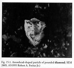
Figure 15.1 Arrowhead-shaped particle of pounded diamond; SEM 200X
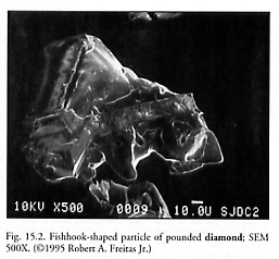
Figure 15.2 Fishhook-shaped particle of pounded diamond; SEM 500X
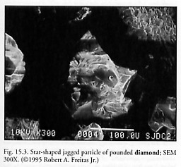
Figure 15.3 Star-shaped jagged particle of pounded diamond; SEM 300X
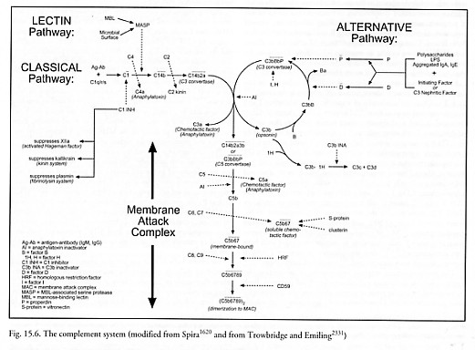
Figure 15.6 The complement system
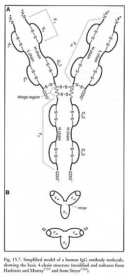
Figure 15.7 Simplified model of a human IgG antibody molecule, showing the basic 4-chain structure
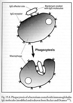
Figure 15.8 Phagocytosis of a bacterium coated with immunoglobulin IgG molecules
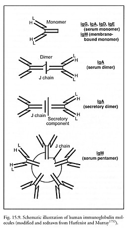
Figure 15.9 Schematic illustration of human immunoglobulin molecules
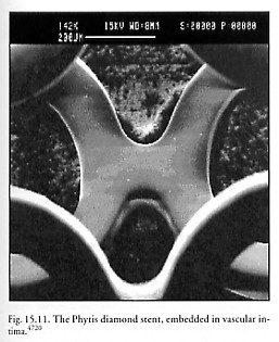
Figure 15.11 The PHYTIS diamond stent, embedded in vascular intima
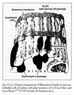
Figure 15.12 Unique arrangement of filamentous bands in sinus endothelial cells of splenic red pulp
Last updated on 30 April 2004