Nanomedicine, Volume IIA: Biocompatibility
© 2003 Robert A. Freitas Jr. All Rights Reserved.
Robert A. Freitas Jr., Nanomedicine, Volume IIA: Biocompatibility, Landes Bioscience, Georgetown, TX, 2003
15.4.2.1 Geometrical Trapping in Lung Vasculature
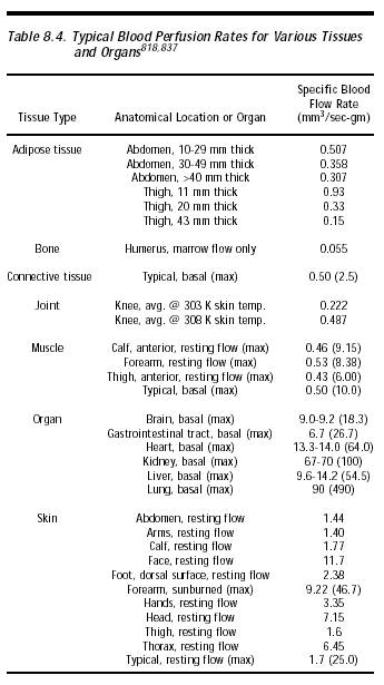
The opportunity for nanorobot trapping via simple geometrical filtration is significant because the lung has the highest specific blood perfusion rate of any organ, typically 90 mm3/sec-gm (~4500 cm3/min) up to a maximum of 490 mm3/sec-gm (~24,000 cm3/min) (Table 8.4). Following IV injection, venous blood flows directly to the heart (Section 8.2.1.1), so the first capillary bed normally encountered by bloodborne injecta is in the lungs. Certainly the injection of large 77- to 125-micron [2683] or 200-micron glass beads [2684] causes vascular embolization. But in general all IV bloodborne particles >7-8 microns are preferentially trapped in the pulmonary capillary bed [2685, 2764] (when this is the first capillary bed through which the bloodborne particles must pass). For instance, 7.4-micron and 11.6-micron diameter polystyrene microspheres administered IV are filtered out by the pulmonary capillary network, mostly during the first pass, with no hemodynamic effect [2679, 2686]. Radiolabeled microspheres administered intravenously to beagle dogs showed that 8- to 25-micron spheres stay in the lung at least 1 month [4495]. 3-micron spheres are rapidly cleared from the lung (most having left after 2 hours [2679]) and are found in liver and spleen after 1 month [4495, 4498]. This effect has long been exploited in radiodiagnostic imaging using albumin microspheres and in the delivery of anticancer agents [2687]. Some large particles, depending on their reactivity, can cause pulmonary granulomas [2685].
Inert particles smaller than 7 microns in diameter can pass through the lung capillary bed without being trapped unless they are aggregated or are very hydrophilic, in which case the pulmonary bed deposition of somewhat smaller particles can be significant [2688]. For example, some 1- to 2-micron diameter engineered liposomes containing negatively charged amphiphiles have optimal deposition in the lung [2689]; accumulation at extrahepatic sites such as the lungs is influenced by liposome size, charge, and composition [5692].
Last updated on 30 April 2004