Nanomedicine, Volume IIA: Biocompatibility
© 2003 Robert A. Freitas Jr. All Rights Reserved.
Robert A. Freitas Jr., Nanomedicine, Volume IIA: Biocompatibility, Landes Bioscience, Georgetown, TX, 2003
15.4.3.3.3 Clearance of Inhaled Particles
The possible mechanical toxicity of particle inhalation, normal environmental dust levels, the clearance of particles from the lungs via the mucociliary escalator (Section 8.2.2), and the role of alveolar macrophages in respect to crystalline particles, especially diamond, has been described in Section 15.1.2. To review, most micron-size particles (similar in diameter to proposed medical nanorobots) that reach the alveoli are quickly cleared by the mucociliary escalator [3064-3070]. This process of clearance is influenced by particle surface chemistry [3087] and by total particle surface area [3088]. Pure mucociliary particle transport has a mean half-life of 2-3 hours [3089], which can be slightly accelerated by oral, or IV, administration of aminophylline [3090]. Particles cleared in this manner are swallowed and exit the body through the alimentary canal unless they are reabsorbed in the gut (Section 15.4.3.3.2) or stomach (which in the case of coal dust particles can lead to an increased risk of gastric cancer [3091]).
Those microparticles not immediately cleared by the escalator are ingested by phagocytes, mostly the pulmonary alveolar macrophages (PAMs) [3092-3094] residing in the alveolar airspaces. (Lavages typically reveal a total of ~109 macrophages present in the human lungs [3095] and a burden of resident particles 0.5-1.2 microns in size [3096, 3097]; in the non-exposed lung, 1-2 macrophages reside in each alveolus in a near-sterile environment [6061].) This process is also, in part, a function of both chemical and physical particle surface properties [767, 3098, 3099], though no comprehensive analysis has yet been done (which will be essential for serious nanorobot design).
Fibroblasts [3103] and leukocytes [766, 3104, 3105] can become involved in clearance as well. In one experiment [3104], intratracheal instillation of rat lungs with 0.5 x 10⁹ microspheres caused an influx of PMN leukocytes from tissues into the pulmonary airspaces. Nevertheless, after 1 day, 77% of the microspheres recovered in bronchoalveolar lavage fluid had been ingested by pulmonary alveolar macrophages and only 19% by PMNs, with 4% of the particles still free [3104]. After 2 days, 95% of the microspheres were inside the macrophages, and ~100% were still present after 4-7 days [3104]. After particle internalization, macrophages generally exit the lungs either: (1) by migrating to the nearest bronchiole and availing themselves of the mucociliary escalator [179, 3099-3102], or (2) by passing into the interstitium (or in the case of interstitial macrophages, accumulating interstitial-resident particles [3106]) and exiting via the blood vessels or lymphatics, often accumulating in regional lymph nodes [3071-3076]. Alveolar macrophages can ingest 1.5-micron diameter glass fibers that are up to 5 microns long, but not fibers that are 60 microns in length [758, 2493]. Fiber inhalation can affect the subsequent lung clearance of microspheres [3107].
How fast are the lungs normally cleared of particles? In a series of studies by Falk et al [3108, 3109], 6-micron monodisperse chemically-inert Teflon particles were inhaled slowly (depositing in small ciliated airways) or normally (depositing in large bronchi and alveolar region) by healthy nonsmokers. About 60% of the particles deposited in the conducting airways during the slow inhalation were cleared after 24 hours. Of the remaining particles, 35% cleared with a half-life of 3.6 days and 65% with a half-life of 170 days [3108]. After the normal inhalation, 14% of the particles retained after 24 hours cleared with a half-life of 3.7 days and 86% cleared with a half-life of 217 days [3108]. A related study of Teflon and polystyrene 6.05-micron and 4.47-micron particles also found ~50% clearance in 24 hours [3110].* Investigations by Langenback et al [3064, 3065] of 2.85-micron diameter carbonized insoluble polystyrene particles instilled in sheep lungs found rapid clearance in 44 hours for tracheobronchial deposition via the mucociliary escalator. This included 2-4 hour clearance of particles deposited in bronchi down to 1 mm in diameter, with slower mostly alveolar clearance over next 30 days. Alveolar deposited particles were sequestered by macrophages and there was no interstitial penetration by alveolar-deposited particles. Macrophages engulfing these particles at low particle burden per cell normally travel only in one direction, from interstitium to alveolus and then to the escalator [3065]. Clearance efficiency generally increases with increasing particle size [3112]. Numerous mathematical models of lung clearance as a function of particle size have been devised [3078-3086]. Clearance of carbon particles from the lung is briefly described in Section 15.3.3.5.
* An older and apparently inconsistent study by Snipes et al [3111] of 3-, 7-, and 13-micron polystyrene microspheres instilled in the lungs of beagle dogs reported that only 2-3% of all particles cleared in a few days via the mucociliary escalator. Of the remaining particles, 3-micron microspheres cleared with a retention half-life of 820 days while the 7- and 13-micron microspheres cleared with a half-life in excess of several thousand days [3111].
Phagocytes may also transport particles from lungs to lymph nodes for presentation to T lymphocytes [3070-3077]. For example, fused aluminosilicate microparticles injected into beagle dog lungs were translocated by alveolar macrophages to left mediastinal, left and right tracheobronchial (TBLN), and left middle and right middle TBLN lymph nodes [3005]. Microparticles of calcium tungstate sprayed into dog lungs are carried by pulmonary macrophages to the regional lymph nodes, with the first particles arriving after 24 hours and peak arrivals at 7 days [3113]. Neutrophils in dogs can make similar journeys [3114]. Lymph nodes may become enlarged due to the arrival of particle-laden alveolar macrophages and particles phagocytosed in resident hyperplastic histiocytic cells [3115].
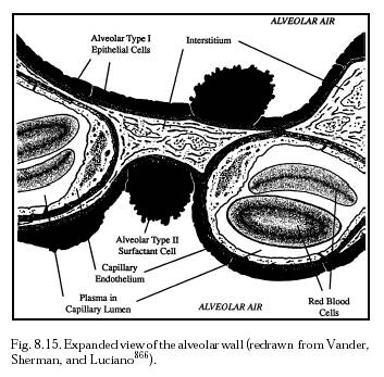
A small amount of particulate matter escapes primary phagocytosis by the alveolar PAMs and PMNs and penetrates the respiratory epithelium, lodging in the interstitium (Figure 8.15) between cells [3116]. In one experiment, 12 hours after carbon particle overloading in mouse lungs some free carbon crossed the type I cells to reach the interstitium and was later observed in peribronchial and perivascular interstitial cells [772]. This triggered a proliferative burst among free interstitial macrophages [772], which can absorb these particles and transport them back across the epithelium and into the alveolar spaces for removal in the usual manner. Particulate matter that avoids this recovery process is later removed from the interstitium along the lymphatic capillaries (initial lymphatics) [180] to the draining lymph node via the lymphatic circulation, particularly the pleural, hilar, or more distant nodes [3117-3120]. 21-50 nm carbon particles instilled into the nasal mucosa generally cannot pass through the epithelial basement membrane unless inflammatory cells (eosinophils) have preceded them. Even then, inert particles might not penetrate further since the interstitial fluid flows outwardly from the mucosa during allergy [3121].
As noted earlier (Section 15.1.2), 2 hours after exposure ~2% of small alveolar-resident particles may penetrate the airway lining and enter the pulmonary interstitium and the phagocytic vacuoles of lymphatic endothelial cells. At 24 hours, these particles are detected in the peribronchial lymphatics and lymph nodes [173, 180, 766], but overall lymphatic clearance is low [766]. For example, in the study by Snipes et al [3111] cited earlier, 1.7% of 3-micron polystyrene microspheres instilled in beagle dog lungs translocated to tracheobronchial lymph nodes during the 128-day study, whereas only 0.2% of the 7-micron particles and none of the 13-micron particles accumulated in tracheobronchial lymph nodes. A related study [3122] found that 1% of 3-micron latex microspheres inhaled by rats and guinea pigs were translocated from lung to lung-associated lymph nodes, whereas none of the similarly-inhaled 9-micron or 15-micron microspheres were found in these lymph nodes. Up to 6% of very small particles, such as are found in diesel exhaust (typically ~0.2 micron median aerodynamic diameter [5933]), make their way to the mediastinal lymph node in rats after 28 days [3123]. 4% of lung burden in dogs exposed to 1.8-micron coal dust was translocated to the tracheobronchial lymph nodes after ~1 year [3124]. Rat-inhaled cristobalite (silica) aerosol particles accumulate in the mediastinal lymph nodes and thymus [3125], and rat-inhaled coal fly ash particles <2.3 microns in diameter are transported to the bronchopulmonary lymph nodes [3126].
Conventional fiber biocompatibility analysis commonly focuses on particle dose, dimension, and durability, with durability determined by inhalation biopersistence (e.g., fiber retention in lung and clearance half life after 5-day animal exposure) and laboratory dissolution rate (e.g., fiber dissolution rate kdis measured in ng/cm2-hr) [6061].
Last updated on 30 April 2004