Nanomedicine, Volume IIA: Biocompatibility
© 2003 Robert A. Freitas Jr. All Rights Reserved.
Robert A. Freitas Jr., Nanomedicine, Volume IIA: Biocompatibility, Landes Bioscience, Georgetown, TX, 2003
15.5.3.4.2 Nanorobotic Lacerative Vasculopathy
Individual free-floating intravascular nanorobots or vasculomobile individual devices or nanorobotic aggregates may occasionally scratch, scrape, or gouge the vascular luminal surface, causing partial or complete loss of local endothelium (Type II damage), resulting in a form of mechanical vasculitis or capillaritis. Since the typical dimensions of bloodborne nanorobots approximate the endothelial thickness (~0.2-2.0 microns [2752, 5953-5955]), transmural Type III damage to the media is unlikely. Turnover studies of rat endothelium show that: (1) injured endothelium can recover an area one cell wide (~1000 micron2) in ~3 hours [4600], (2) the natural loss rate is ~0.1% of endothelial cell area per day (~1 micron2/day) [4601], and (3) the steady-state vascular denudation area is ~0.125 micron2/cell [4602].
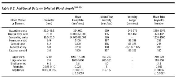
Smooth nanorobot hulls lacking sharp edges or protrusions (during travel), boundary layer effects, and low blood velocity throughout the nonarterial vasculature should ensure that major “sandblasting” type erosion [3921, 3922] is unlikely to occur inside human blood vessels even at the highest nanocrits consistent with continuous flow. Free-floating nanorobots that collide with blood vessel walls (given the no-slip condition at the wall) produce minimal shear forces, on the order of <~0.1 N/m2 (Section 9.4.2.2). This is less than the 1.0-2.6 N/m2 shear forces normally encountered in arteries and capillaries due to normal blood flow and the 0.14-0.63 N/m2 shear forces in veins, but may be sufficient to cause a small biological response from the vascular endothelium (Section 15.5.3.1.1). Applying the maximum bloodstream velocity of ~1 m/sec (Table 8.2) to the impact-scratch relation (Eqn. 9.96) given in Section 9.5.3.6, it is clear that particle-wall collisions should produce only harmless submicron nicks even in the most turbulent arteries.
Nevertheless, some caution is warranted because natural endothelial cell wounding of 1-18% of all cells, possibly erosionally-derived, has been observed in rat aorta [3923]. Erosion of cultured fibroblast monolayers (simulating the vascular endothelium) using MHz ultrasound at acoustic pressures of ~106 N/m2 is enhanced by the presence of a microbubble (particulate) contrast agent [3924]. Injection of crystalloid cardioplegic solutions into canine hearts at pressures >110 mmHg and at peak flow rates >25 ml/sec also causes a higher incidence of mechanical-physical trauma to the vascular endothelium and the endocardium [3925]. In another unusual case, intravenous self-injection by a drug abuser of dissolved tablets containing microcrystalline cellulose as filler material produced numerous microcrystalline cellulose pulmonary emboli, intravascular foreign body granulomas, focal necrosis and edema of the pulmonary parenchyma, and fatal vascular destruction [3926].
Endothelial abrasion alone may not stimulate neointimal thickening [4599] but inevitably must involve some endothelial cell loss [3927] and other biological responses. For example, mechanically scraping cultured endothelial cells causes growth factor to be released within 5 minutes, not abating for at least 24 hours thereafter, due to plasma membrane disruption [3928]. In cases of vascular dissection, a piece of the endothelium peels up (with the break often extending deeper, into the media), making an intimal flap that defines regions of true and false lumina [3837]. Sometimes this may induce an intramural hematoma in the aortic wall [3837]. Endothelial cells mechanically damaged with a razor blade activate extracellular-signal-regulated kinases within ~300 sec, releasing fibroblast growth factor (FGF-2) which in turn induces intimal hyperplasia [3929]. Nanorobots which detect FGF-2 are alerted that mechanical endothelial injury has taken place. By absorbing the cytokine using molecular sorting rotors, the hyperplasia signal could be suppressed by a team of nanorobots, if desired (Section 7.4.5.4). However, shear-induced endothelial denudation of healthy canine arterial endothelium appears not to occur at shear stresses up to at least 200 N/m2 [3930]. The role of erythrocyte collisions with vascular walls on the detachment rate of endothelial cells is just starting to be seriously investigated [3931].
Last updated on 30 April 2004