Nanomedicine, Volume IIA: Biocompatibility
© 2003 Robert A. Freitas Jr. All Rights Reserved.
Robert A. Freitas Jr., Nanomedicine, Volume IIA: Biocompatibility, Landes Bioscience, Georgetown, TX, 2003
15.4.3.2.3 Phagocytosis in Liver Vasculature
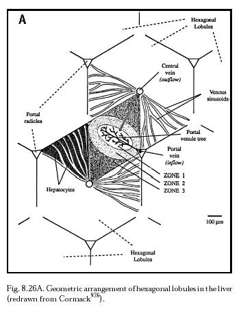
It is well known that if particles consisting of carbon (India ink [777, 2930]) or vital dyes are injected into the blood, the macrophages (Kupffer cells) of the liver (along with the phagocytic cells of the spleen) ingest most of them [2888]. Kupffer cells are ~15- to 20-micron stellate phagocytic cells with a 70 nm thick fuzzy coat including a 15 nm thick glycocalyx [2931], mechanically attached to the sinusoid (Section 15.4.2.2) endothelial cells of the liver. Kupffer cells partially occlude the sinusoid lumen but have no functional attachments to the endothelial cells or to the underlying hepatocytes, and are partially motile [2932]. Their customary positioning, predominantly at the periportal end of the sinusoids, confirms that they monitor arriving blood, looking for particles to remove from the flow [2845]. Distribution in liver lobules (Figure 8.26A) is 43% periportal, 28% midzonal (midacinic), and 29% in the central area (perivenous), with periportal cells larger and more active than central cells [2704]. Kupffer cells can phagocytize particles of dirt, worn-out blood cells including red cells and platelets, and bacteria – and, presumably, inert nanodevices. The cells possess internal inventories of rolled-up spare membrane. This allows a more rapid ensnarement of particulate matter [2932]. Kupffer cells can internalize 5-micron diameter IgG-coated sheep erythrocytes (SRBCs) at a peak rate of 3-4 SRBCs/minute, and C3b complement-coated SRBCs at a peak rate of 5-6 SRBCs/minute [2845].
The Kupffer cell population constitutes ~31% of liver sinusoidal cells [2704], with a mean of 14-20 x 106 cells/gm liver tissue [2704] or ~25 billion Kupffer cells in the entire organ [2845, 2933]. Long-term observations reveal that the mean number and distribution of cells is unchanged over 3 months, indicating that these resident macrophages represent a long-living (i.e., many months) and self-renewing population. Population turnover is slow, with a cell-cycle of ~52 hours including an S-phase of ~7 hours [2934]. About 75% of Kupffer cell population growth comes from cell replication, while the remaining 25% of population growth results from extrahepatic recruitment of macrophage precursors [779, 2930, 2934].
A colloidal particle that undergoes opsonization is usually removed rapidly and efficiently by liver macrophages [2904]. The particles are trafficked to the lysosomal compartment of the cell where a battery of enzymes can degrade labile structures [2904]. The half-life of the uptake process can be less than 1 minute for small (e.g., <100 nm) opsonized particles, with more than 90% of the administered colloid being sequestered inside the cells [2682].
The first nanorobotic line of defense against Kupffer cell phagocytosis is to reduce or to avoid opsonization by blood proteins that would make nanorobots visible to the RES. It may be possible to devise proteophobic coatings (Section 15.2.2.1) to accomplish this objective. For example, particulate matter not coated with blood proteins does not adhere to the 70-nm fuzzy coat lining the cells of hepatic sinusoids [2935]. Under normal conditions, formed elements of the blood and lipid droplets like chylomicrons also do not adhere to the wall of sinusoids [2935]. If diamondoid surfaces can be rendered unwetted or unwettable by fibrinogen and other blood proteins, or can be appropriately masked or pegylated [2904] (Section 15.2.2.1), then physically intact nanorobots might not be efficiently recognized and phagocytosed by the Kupffer cells or the other phagocytic cells of the liver (see below).
If nanorobots are recognized as foreign objects suitable for phagocytosis, uptake and distribution of nanorobots will depend to some degree upon their size. Studies of uptake and distribution as a function of particle size have been done. In one experiment [2936], polystyrene microspheres 0.05 microns and 0.5 microns in diameter were administered IV to rats. Both particle sizes were mostly distributed to the liver, with small but significant amounts distributed to lung (in the case of the 0.05-micron particles) and spleen (for the 0.5-micron particles). In the liver, uptake of the smaller 0.05-micron particles went 59% to Kupffer cells, 28% to parenchymal cells (e.g., hepatocytes), and 13% to endothelial cells; uptake of the larger 0.5-micron particles went 71% to Kupffer cells, 24% to endothelial cells, and only 5% to parenchymal cells [2936]. Passively circulating inert nanorobots would likely be similarly distributed.
Another experiment [2836] using chemically inert latex particles also found that uptake by a particular phagocytic cell type was determined by particle size. Sinusoidal endothelial cells can internalize particles up to 0.23 microns in size under physiological conditions in vivo, while larger particles normally are taken up by Kupffer cells. However, when Kupffer cell uptake is impaired (e.g., by alcohol), endothelial cells can uptake particles up to 1 micron in diameter after the injection of an excess amount of latex particles [2836]. Splenic macrophages (Section 15.4.3.2.4) can also do this [2937]. Endothelial cells thus constitute a second line of defense in the liver, removing foreign materials from the blood when Kupffer cell phagocytic function is totally disturbed. The total cellular plasma membrane surface area of each cell type, per cm3 of liver parenchyma, is 1160 cm2 for hepatic sinusoidal endothelial cells and 325 cm2 for Kupffer cells [2938].
A similar study [2939] of the endocytosis of latex particles 0.33-, 0.46-, and 0.80-micron in diameter by sinusoidal endothelial and Kupffer cells in rat liver found that after 10 minutes all three sizes were incorporated by the luminal cell surface of the perikarya or thick portion of the endothelial cells in vitro (bicarbonate-perfused liver) but in numbers far less than in the Kupffer cells. However, in vivo endocytosis of these particles was observed in Kupffer cells but not in endothelial cells. A particle ingested by an endothelial cell was surrounded by a large patch of bristle coat, whereas in Kupffer cells the particle was engulfed by the ruffled membranes or sank into the cytoplasm without a large patch of bristle coat, suggesting different endocytotic mechanisms for the two cell types [2930, 2939].
If Kupffer cells cannot break down ingested particles – as would most likely be the case for diamondoid medical nanorobots – what is the ultimate fate of these cells? (For the fate of phagocytosed nanorobots, see Section 15.4.3.6.) Fujita and colleagues [777] studied the long-term changes in Kupffer cells in mice that were given intravenous India ink. Aggregates of Kupffer cells containing many vacuoles stuffed with 10-100 nm carbon particles appeared in the sinusoidal lumen, Disse space and interlobular connective tissue space 3-4 days after ink injection. After 1 month, large clumps of aggregated Kupffer cells containing numerous carbon-filled vacuoles up to 9 microns in diameter were distributed in the Disse space and other connective tissue, with cells in close contact and partly fused with one another. After 3-6 months, large multinucleate foreign body giant cells (Section 15.4.3.5) with numerous large vacuoles containing densely-packed ink particles were visible throughout the liver tissue, probably formed by fusion of particle-stuffed Kupffer cells. Some endothelial cells also stored ink particles in cytoplasmic vacuoles for as long as 6 months after injection [777]. Particle doses were evidently too low to produce clinically observable pathological effects on mouse liver function.
In a subsequent experiment by the same group [2940], mouse Kupffer cells in vivo took up 0.2- and 2.0-micron polystyrene latex particles and most cells were stuffed with the particles after 2 days. After 1 month, large Kupffer cell clumps or aggregates (which the researchers called granulomas) 80-120 microns in diameter were observed in the liver connective tissue spaces (i.e., Disse, interlobular and subperitoneal) composed mostly of cells heavily laden with latex particles. Hepatic sinusoidal endothelial cells, attenuated in shape, also took up 0.2-micron particles (but only very rarely a 2-micron particle) into their cytoplasm [2940]. After 8 months, numerous large granulomas were distributed throughout the liver in the interlobular or subperitoneal connective tissue spaces [2940].
These results suggest that medical nanorobots should be designed first to avoid recognition and uptake by phagocytic cells, and second to actively escape (Section 15.4.3.6) from such cells during or after hepatic phagocytosis, in order to forestall significant foreign body giant cell and granuloma formation (Section 15.4.3.5) in the liver, or unintentional RES blockade (Section 15.4.3.6.10).
Last updated on 30 April 2004